Figure 3.
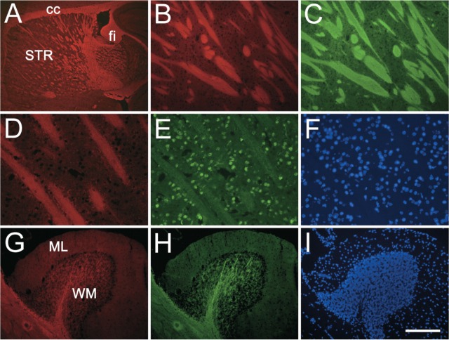
NIM-1 staining in the normal mouse brain. Para-sagittal sections were stained with NIM-1 (red, A, B, D, G) costained with either myelin basic protein (MBP) (green, C, H) or NeuN (green, E), and mounted with the nuclear stain DAPI (blue, F, I). NIM-1 stains distinct white matter structures in the brain (A), such as striatum (STR), corpus callosum (cc), and fimbria (fi). In the striatum (B-F), NIM-1 stains fiber bundles (B), completely overlapping MBP staining (C). The compatibility of NIM-1 staining (D) with other fluorescence staining is further demonstrated by NeuN immunostaining (E) and nuclear stain DAPI (F). In the cerebellum folium, NIM-1 stains the white matter (WM), colocalizes with MBP (H), while leaving molecular layer (ML) unstained. Bars: A = 500 μm; B, C = 100 μm; D-F = 50 μm; G-I = 200 μm.

