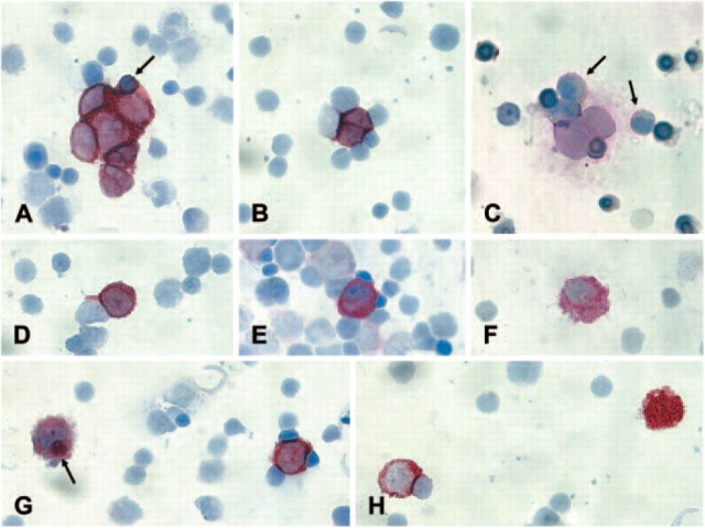
Figure 1.
Immunocytochemical analysis of bone marrow (BM) slides from neuroblastoma (NB) patients according to the standardized protocol. (A) CPCs (criteria-positive cells) forming a clump. NB cells in clumps do not always display a round nucleus because they adjust their form to the clump. The entrapped erythroblast (arrow) is smaller than the NB cells and displays a different chromatin structure. (B) The two CPCs mold in a different way compared with the surrounding hematopoietic cells. (C) Two CPCs showing the typical nuclear size, chromatin structure, and nuclear/cytoplasmic ratio that clearly differs from hematopoietic cells. The myeloid cells (arrow) are passively stained because of the shedding of the GD2 antigen by the NB cells. (D) If the morphological and immunological criteria are fulfilled, even a single cell can be identified as CPC. (E) The staining intensity of this cell is comparable to that of a CPC but the nuclear shape, size, chromatin structure, and the nuclear cytoplasmic ratio do not fulfill the criteria. Therefore, the cell is classified as not convincingly interpretable cell. (F) Neither the nuclear/cytoplasmic ratio nor the vesicular structure of the cytoplasm fulfills the criteria. The cell has the cytological features of a histiocyte. (G) The cell on the left side has the same size and displays the same staining intensity as the CPC on the right. However, the nuclear shape and the nuclear/cytoplasmic ratio do not fulfill the criteria. Moreover, the cell contains GD2-positive material in the cytoplasm (arrow). This is a typical feature of a macrophage. (H) Strongly positive material (right) without a visible nucleus is not reported. CPC on the left.

