
Figure 3.
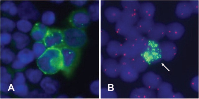
Automatic immunofluorescence plus FISH allows the sequential immunological staining and molecular cytogenetic characterization of disseminated neuroblastoma cells. (A) GD2-positive cells (green fluorescence). The nucleus is stained with DAPI (blue fluorescence). (B) The genetic makeup of the GD2-positive cells is visualized by FISH. Only one GD2-positive cell displays MYCN amplification (green fluorescence signals). GD2-positive cells displaying the same genetic aberrations as the primary tumor are called FISH-positive cells.