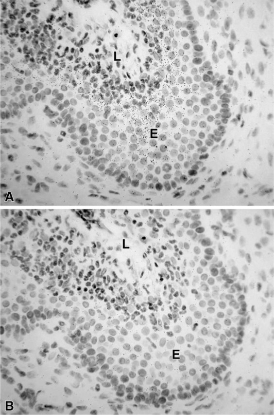
Figure 4.

(A) Micrographs illustrating the hybridization signal obtained in the epithelial cells (E) of the uterine cervix. (B) Consecutive section hybridized with the sense probe. No accumulation of silver grains can be detected. L, lumen. Exposure time 21 days. Original magnification ×580.
