
Figure 5.
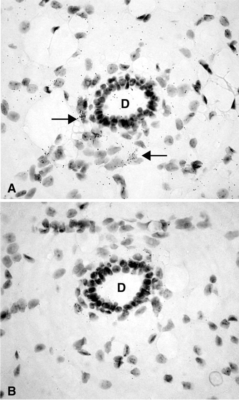
(A) Representative micrograph illustrating the hybridization signal obtained in a mammary gland of a female mouse. Labeling can be observed only over a few stromal cells (→) in proximity to an unlabeled duct (D). (B) Consecutive section hybridized with the sense probe. Only diffuse background can be observed. Exposure time 45 days. Original magnification ×700.