
Figure 1.
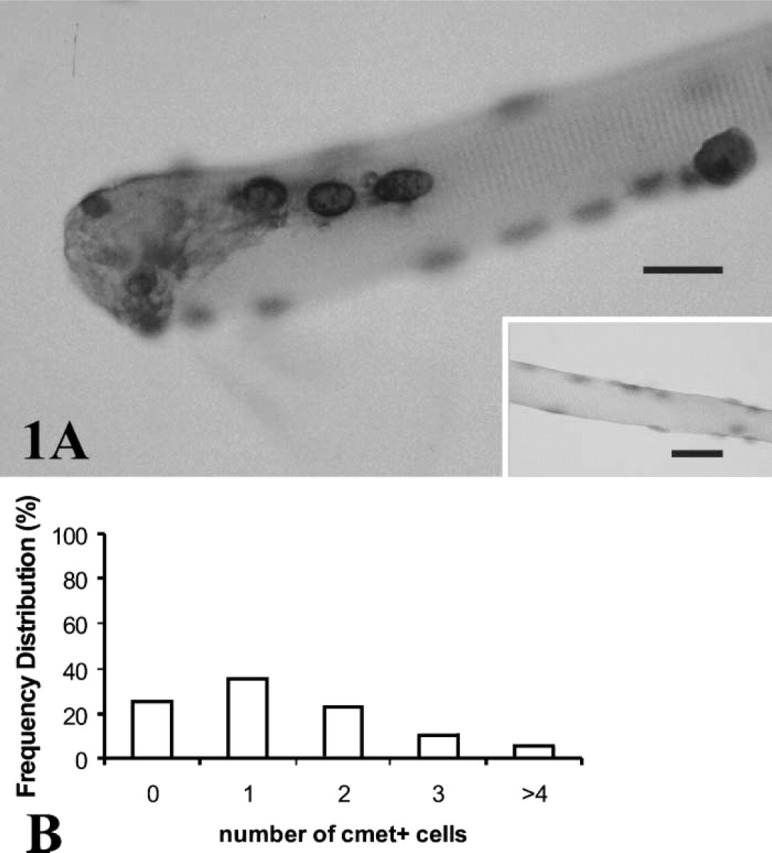
(A) Satellite cells were identified on single fibers using ISH and riboprobes to detect c-Met mRNA. Four cells appear on this fiber, as detected by a dark rim of cytoplasm surrounding paler nuclei, lightly counterstained with hematoxylin. Fiber cytoplasm did not stain for transcripts of c-Met. (Inset) Negative control for the in situ procedure omitting the riboprobe for c-Met shows no signal for mRNA. (B) Frequency distribution [proportion (%) of total fibers counted] of quiescent satellite cells per fiber that were positive for c-Met mRNA in a population of 618 normal mouse FDB fibers, fixed 18 hr after isolation and without stretching. From 0 to 6 cells per fiber expressed c-Met transcripts, with a mean (± SE) of 1.38 ± 0.07 c-Met+ cells/fiber.