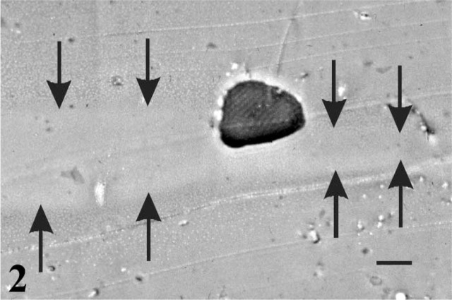
Figure 2.
Light micrograph of an isolated fiber in transverse section partially embedded in the Vitrogen layer. Sections through all layers were made perpendicular to the original culture plate (2 μm in thickness) and stained with toluidine blue to show the fiber (dark) and the paler Vitrogen layer (delimited by arrows) above the region of resin that replaced the flexible substrate (lower gray region). Note the variation in the thickness of the Vitrogen layer. Bar = 20 μm.

