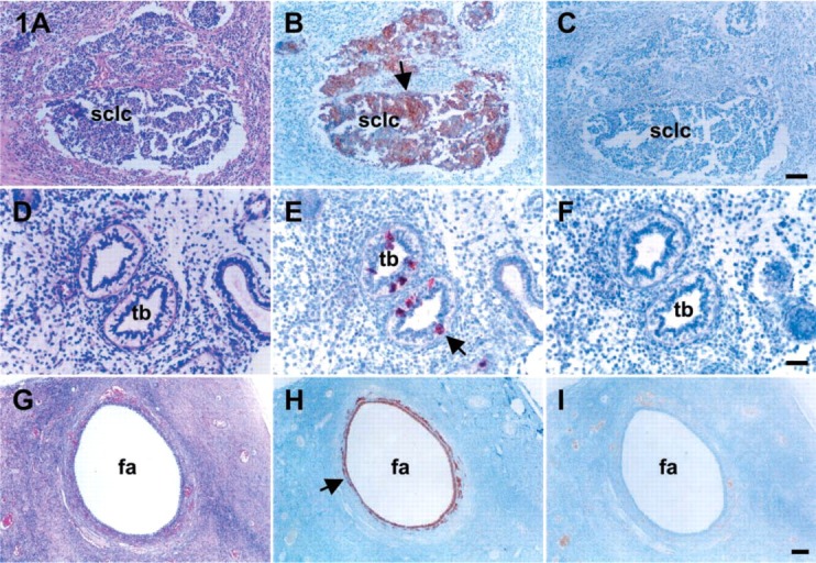
Figure 1.

IHC for c-kit, SCF, and inhibin-α in the present experiments was validated using tissues from small-cell lung carcinoma (SCLC) 13-week-old fetal lung, and noninvasive normal ovary. With hematoxylin and eosin (A,D,G), the tissues were localized by IHC for c-kit (B), SCF (E), and inhibin-α (K). The IHC localization of c-kit was identified in SCLC. SCF was localized in the epithelium of terminal bronchi. Immunoreactivity for inhibin-α was localized in the granulosa and theca cells of antral follicles in a 27-year-old human ovary. (C,F,I) Negative controls performed without primary antibodies represent the validities of the present immunohistochemistries. Arrows indicate the immunoreactive localization of c-kit (B), SCF (E), and inhibin-α (H). sclc, small-cell lung carcinoma; tb, terminal bronchi; fa, follicular antrum; SCF, stem cell factor. Bars: A-C = 100 μm; D-F = 50 μm; G-I = 200 μm.
