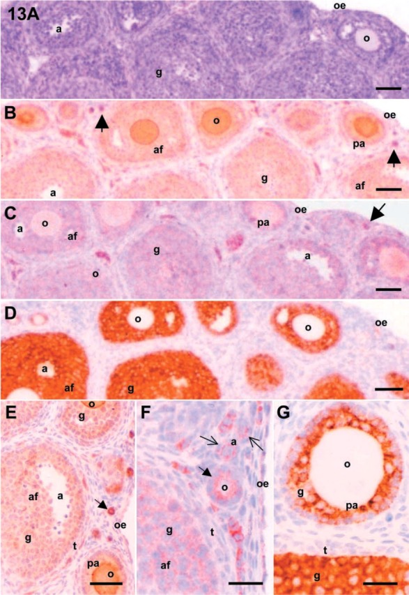
Figure 13.

Microphotographs of mouse ovaries. Serial sections from the immature (21 dpp) mice ovaries were stained with hematoxylin and eosin (A) and immunoreactive antigens for c-kit (B), SCF (C), and inhibin-α (D) were localized. The localizations of antigens for c-kit, SCF, and inhibin-α were also shown in panels E-G, respectively. Abbreviation: a, antrum; af, antral follicle; g, granulosa cell; o, oocyte; oe, ovarian epithelium; pa, preantral follicle; t, theca cell. Arrows indicate primordial follicles. Bars: A-D = 50 μm; E = 100 μm; F,G = 25 μm.
