
Figure 4.
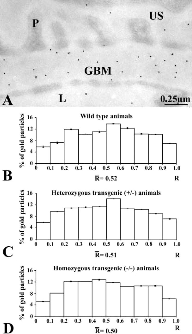
Immunocytochemical localization of laminin. (A) Immunogold labeling of an entactin-1-null heterozygous animal tissue. The gold particles revealing laminin antigenic sites are restricted to the GBM with a rather homogeneous distribution across the GBM. (B-D) Histograms of the distributions of laminin labeling across the GBM of the wild-type (B), heterozygous (C), and homozygous (D) animals. The three histograms display similar distributions, with no changes among the groups of animals.