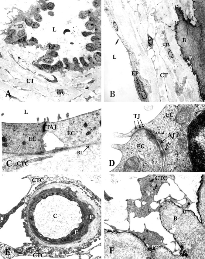
Figure 4.

Transmission electron microscopy of the endolymphatic duct (ED) epithelium and the periductal connective tissue (CT). EP, ED epithelium; L, lumen of the ED; CTC, connective tissue cells. (A) Folded epithelium with basal cell processes extending into the surrounding CT, in the intermediate part of the human ED. (B) Flat epithelium with underlying CT in the proximal part of the human ED. Note the interconnecting CT cells of the periductal tissue. B, bone matrix. (C) Flat epithelium with apical microvilli (MV), and intercellular tight and adherens junctions (TAJ). EC, epithelial cell; BL, basal lamina. (D) High-power transmission electron microscopy of intercellular contacts between the epithelial cells. TJ, tight junctions; arrows show three junctional strands; AJ, adherens junctions. (E) A capillary of the periductal CT. E, endothelial cell; P, pericyte; C, lumen of capillary. (F) A CT cell making contacts with the bone matrix (B) and adjacent cell.
