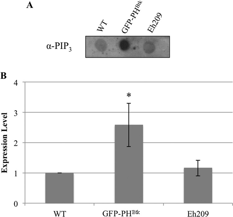
FIG 7.

GFP-PHBtk overexpressors display altered PI(3,4,5)P3 levels. Phosphoinositides were extracted from whole-cell lysates, and PI(3,4,5)P3 levels were measured using dot blots with antibodies specific to PI(3,4,5)P3. (A) A typical dot blot is shown. (B) Blot densities were analyzed by ImageJ software (version 1.42q). The density for the untransfected wild type (WT) was arbitrarily set to 1, and all other data are reported as a ratio to the WT. The data represent the means ± SD from 3 trials. PI(3,4,5)P3 levels were higher in GFP-PHBtk-expressing cells (*, P < 0.05) than in WT cells or in a control cell line expressing an irrelevant protein, luciferase (Eh209).
