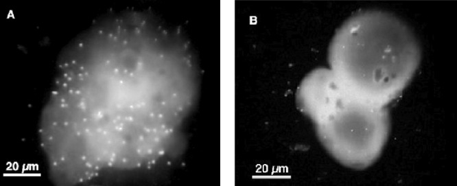
FIG 2.

Representative fluorescence microscopy images of V. vulnificus E-genotype (A) and C-genotype (B) cells attached to a single chitin magnetic bead. Image color, contrast, and brightness were applied to each image by using the Macintosh Preview application.
