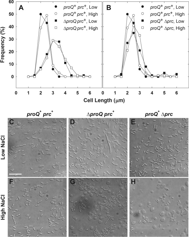
FIG 2.
Impacts of proQ and prc lesions on bacterial morphology. Bacteria were cultivated in MOPS medium to late exponential phase as described for transport assays (25) and visualized by DIC microscopy (see Materials and Methods). (A and B) Media were unsupplemented (0.25 mol kg−1; closed symbols) or supplemented with 250 mM NaCl (0.75 mol kg−1; open symbols). Length distributions are shown for 100 rod-shaped cells of strains RM2 (circles; proQ+ prc+) and WG1119 (squares; RM2 ΔproQ856::FRT) (A) and RM2 (circles) and WG703 (squares; RM2 Δprc3::kan) (B). Subsequent experiments revealed that ΔproQ856::FRT renders the bacteria ProQ and Prc deficient (see Fig. 4). For strain WG1119, 3% of measured cells in cultures at low osmolality and 4% of measured cells in cultures at high osmolality were greater than 6 μm in length. No measured cells in cultures of strain RM2 or WG703 at low or high osmolality were greater than 6 μm in length. (C to H) Representative DIC micrographs are shown for strains RM2 (C and F), WG1119 (D and G), and WG703 (E and H) cultivated in unsupplemented medium (low NaCl; C, D, and E) or NaCl-supplemented medium (high NaCl; F, G, and H). Spherical cells with highly refractive, crescent-shaped internal structures are evident in panels G and H. (In Fig. S1 in the supplemental material, the internal structures are more evident in the corresponding panels [E and F].) Bars, 10 μm.

