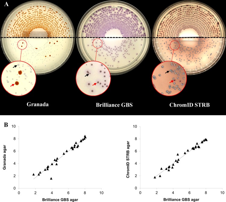
FIG 1.
Comparison of three chromogenic media for GBS screening. (A) Pictures of Granada (left), Brilliance GBS (middle), and ChromID STRB (right) agar plates inoculated with two samples yielding averages of 6.42 × 10⁷ CFU/ml (top) and 7.70 × 10² CFU/ml (bottom). In the enlarged parts of the picture, red arrows indicate target-colored colonies of S. agalactiae and black arrows non-GBS colonies. Agar plates were inoculated using the EasySpiral Dilute instrument and photographed with the Scan 1200 reader (see the text for details). (B) Correlations of GBS loads in 29 samples recovered on Granada and Brilliance GBS media (left graph) (Pearson coefficient of 0.97, P < 0.0001) and on ChromID STRB and Brilliance GBS media (right graph) (Pearson coefficient of 0.98, P < 0.0001).

