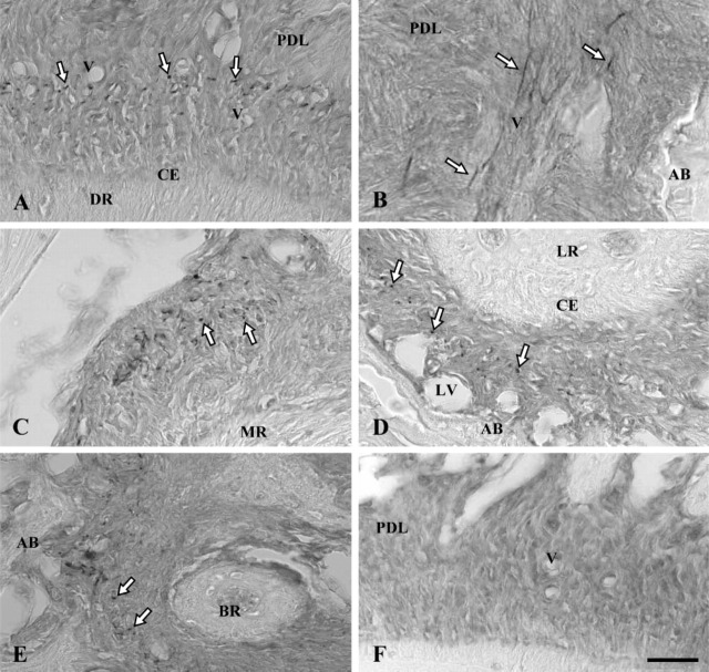
Figure 1.
Immunoperoxidase staining of rat molar periodontal ligament for elastin (A-E) with control section (F). AB, alveolar bone; CE, cellular cementum; PDL, periodontal ligament; V, small blood vessels; LV, large blood vessels. (A) Distal side of periodontal ligament of distal root (DR) sectioned horizontally. Dot-like stains (white arrows) are concentrated mainly in the central zone of the ligament where small blood vessels are localized. (B) Elastin-positive fibers (white arrows) are oriented in the apico-occulusal direction along blood vessels. (C) Stained dotlike structures (white arrows) are concentrated on the distal side of the periodontal ligament of the mesial root (MR). (D) On the proximal side of the ligament of the lingual root (LR), dot-like stains (white arrows) are distributed in the central zone of the periodontal ligament but not around large blood vessels localized in areas close to alveolar bone. (E) Immunoreaction products (white arrows) are present in the periodontal ligament around the apex of the buccal root (BR). (F) Control section in which no immunoreaction product is present. Bar = 80 μm.

