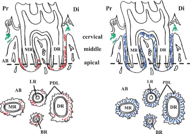
Figure 3.
Schematic drawings of distribution of elastin and fibrillin in the periodontal ligament of rat mandibular first molars. Top: Sagittal section of roots of first molar. Bottom: Section along the horizontal plane at a level indicated by the broken line in the top drawing. Elastin (left, indicated in red) is mainly concentrated in the apical region of the periodontal ligament. Fibrillin (right, indicated in blue) is more widely distributed throughout the ligament. Elastin of gingival connective tissue is indicated in green. AB, alveolar bone; PDL, periodontal ligament; DR, distal root; MR, mesial root; LR, lingual root; BR, buccal root; Di, distal side; Pr, proximal side.

