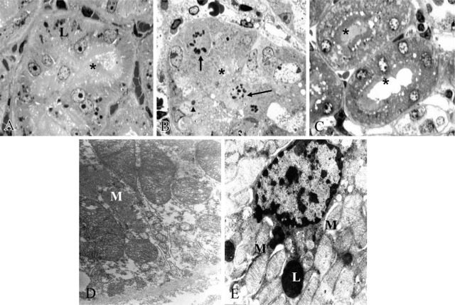
Figure 7.
Toluidine-blue-stained semithin sections. Mitotic figures (arrows) and necrotic nucleus (arrowhead) and detached brush border (∗) were present in the mercury-treated group (B) but were absent in the MEL-treated group (A) and after mercury + MEL (C). Ultrastructure of straight proximal tubule treated by mercury (D) displayed altered mitochondria but recovered after MEL supply (E). Lysosome (L), Mitochondria (M).

