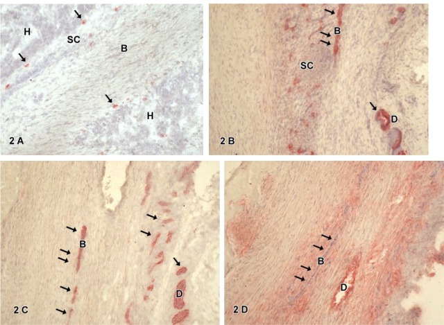
Figure 2.
(B-D) Immunohistochemistry for hepatic marker CK-18 (B), biliary marker CK-7 (C), and stem cell marker Thy1 (D) of mixed type hepatoblastoma with mainly fetal cells. CK-18 staining (B) shows single positive biliary cells (arrows) embedded by small cells (SC) not positive for CK-18. CK-7 expression (C) is seen in aberrant bile ducts (B, arrows) or atypical biliary ducts (D). Thy1-positive cells (D) are found in atypical ducts (D); note that fetal biliary cells (B) do not express Thy1 (arrows). The connective tissue showed a diffuse staining in the Thy1 immunocytochemistry.

