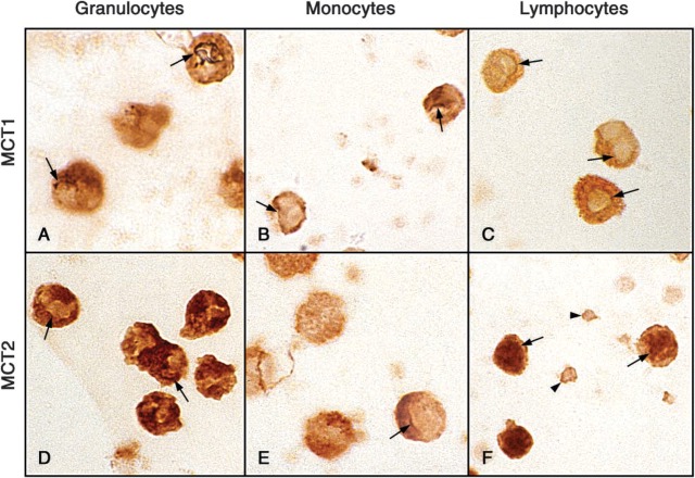
Figure 6.
Evidence for nuclear envelope staining by MCT1 and MCT2 antibodies in each of the three leukocyte fractions at high magnification (×1615). In each panel, the arrows touch a curvy line at nuclear folds or perimeter, which is darker than the nucleoplasm or cytoplasm on either side; this is our requirement for a convincing indicator. In F, the arrowheads point to two platelets with plasmalemmal staining.

