
Figure 5.
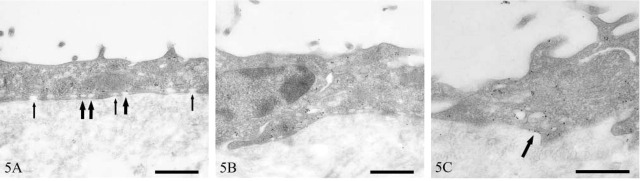
Transmission electron micrographs of human peritoneal mesothelium immunostained for caveolin-1. Immunopositive (thick arrows) and immunonegative (thin arrows) vesicles (A), clustering of immunopositive vesicles around an intercellular junction (B), and linear plasmalemmal staining (arrow) (C). Bars = 500 nm.