
Figure 4.
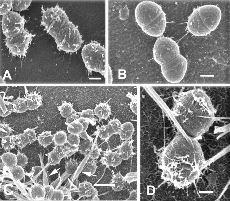
(A) Low-voltage SEM of E. faecalis biofilm. E. faecalis cell fixed in aldehyde cocktail containing 0.15% ruthenium red. Finger-like projections extend outward from the glycocalyx. Bar = 200 nm. (B) Fixation of E. faecalis in aldehyde containing 0.0075% lysine. Short fibrils anchor cells to the substratum, but only sparse fibrils can be seen on the glycocalyx. Bar = 200 nm. (C) Low magnification of E. faecalis biofilm fixed in aldehyde containing 0.15% alcian blue and 0.15% safranin O. Glycocalyx consists of short filaments protruding from the cell surface and large, thick, curved rods (arrows). Bar = 1 μm. (D) Higher magnification of glycocalyx from cells fixed as in C, showing the origin of large, curved rods directly at the cell surface (arrowhead). Short fibrils can be seen on both glycocalyx and substratum. Bar = 200 nm.