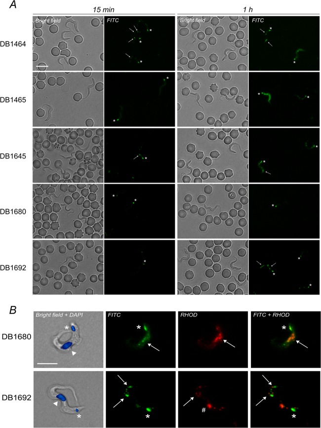
FIG 1.
(A) Fluorescence images of infected rat blood incubated ex vivo with the five green fluorescent diamidines (note that the parasite position can change between the bright-field and corresponding fluorescence micrographs), 40× objective. (B) In vitro trypanosomes treated with DB1680 and DB1692 (50 μM, 1 h, 37°C), 100× objective; 4′,6-diamidino-2-phenylindole (DAPI) (20 μM) was used as a DNA counterstain. Asterisk, kinetoplast; arrow, cytoplasmic corpuscles, possibly acidocalcisomes; arrowhead, nucleus; hash mark, possibly lysosome. Bar, 10 μm.

