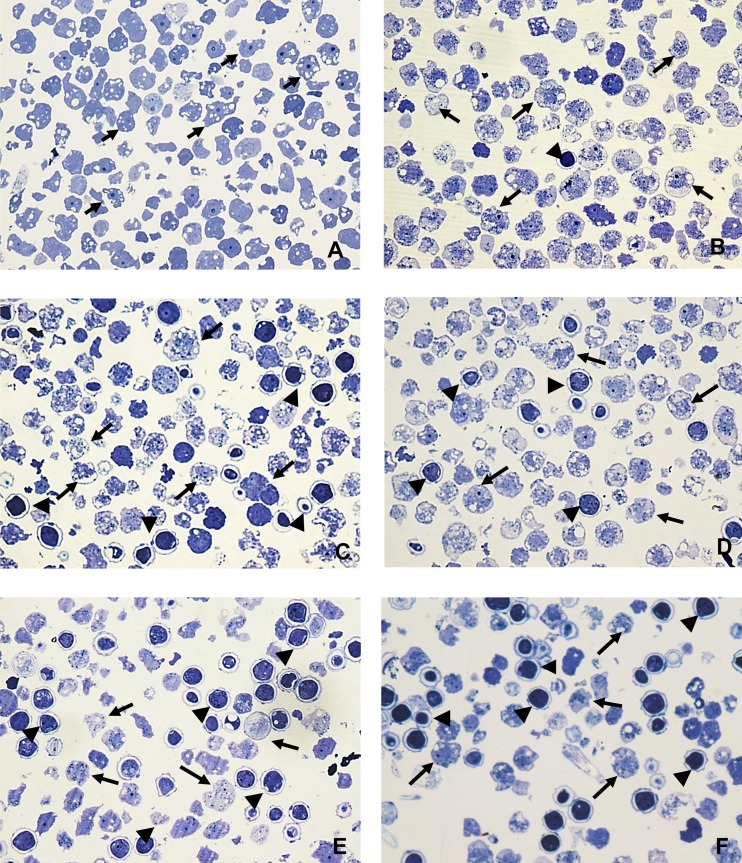
FIG 3.
Light microscopy of epoxy-embedded semithin sections, stained with toluidine blue, of A. castellanii trophozoites incubated in the presence and absence of corifungin. Trophozoites were incubated with 200 μM corifungin for different time periods. (A) Amebae (arrows) with fresh medium only. (B) Amebae incubated with 200 μM corifungin for 24 h. Arrows indicate trophozoites, and the arrowhead indicates a cyst. (C) Amebae incubated with 200 μM corifungin for 48 h. The damage in the trophozoites (arrows) is evident, and there was an increase in the number of the cysts (arrowheads). (D) Amebae incubated with 200 μM corifungin for 72 h. More damage is seen in the trophozoites (arrows), and damage is also found in the cysts (arrowheads). (E and F) Amebae incubated with 200 μM corifungin for 96 to 120 h. Cell remains of trophozoites (arrows) and more damage in the cysts (arrowheads) are seen. Magnification, ×60.

