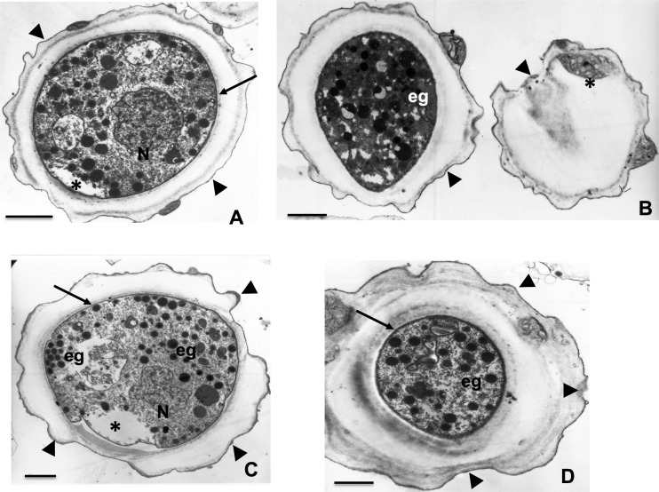
FIG 5.
Transmission electron microscopy of A. castellanii cyst formation after incubation of trophozoites with 200 μM corifungin for 120 h. Encystment of the ameba is evident. Cell wall (arrowheads) is present. Inside the cyst, the trophozoite (arrow) presents signs of damage (*). Electron-dense granules also appear (eg). In many cysts, loss of the inner wall and cell wall disruption are observed (arrowheads). Bars, 2 μm.

