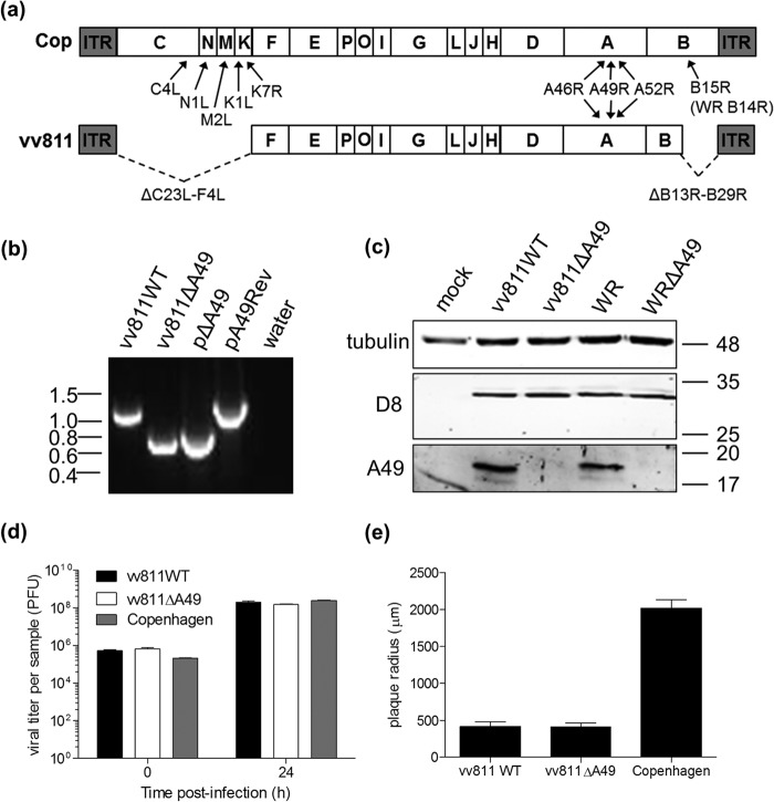
FIG 1.
Construction of vv811 lacking A49R. (a) Schematic of the genome structure of VACV strains vv811 and Copenhagen (Cop), with the position of the known intracellular NF-κB inhibitors downstream of TNF-α or IL-1β indicated. ITR, inverted terminal repeat. (b) The A49R gene from vv811 was deleted by transient dominant selection (see Materials and Methods) to yield vv811ΔA49. A matching wild-type virus (vv811WT) was isolated from the same intermediate virus. The phenotype of the resolved viruses was analyzed by PCR from proteinase K-treated infected BSC-1 cell lysates using primers annealing to the flanking regions of A49R. The resulting PCR product sizes were compared to those obtained from the plasmid templates (pΔA49 and pA49Rev). Molecular mass markers (in kbp) are indicated on the left. (c) Expression of A49 by the recombinant vv811 viruses in lysates of BSC-1 cells mock infected or infected for 16 h with 2 PFU per cell was analyzed by immunoblotting using an anti-A49 polyclonal antiserum and anti-tubulin and anti-D8 antibodies as controls. Molecular mass markers (in kDa) are indicated on the right. (d) A549 cells were infected in duplicate with vv811 recombinant viruses or Copenhagen at 5 PFU per cell. Cells were then harvested at 0 h and 24 h, and the infectious titer of intracellular virus was determined by plaque assay on BSC-1 cells. (e) BSC-1 cells were infected with 50 PFU per well of vv811 recombinant viruses or Copenhagen, and plaques were allowed to form for 5 days. Cells were then stained with crystal violet, and the plaques were imaged and measured. Data are represented as the mean plaque radius (μm) ± SD.

