
FIG 2.
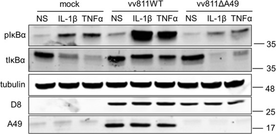
pIκBα does not accumulate in vv811ΔA49-infected cells. A549 cells were mock infected or infected for 16 h with 2 PFU per cell of the indicated viruses and then stimulated for 30 min with IL-1β (25 ng/ml) or TNF-α (100 ng/ml) or nonstimulated (NS) as a control by incubation with medium alone. The cells were lysed under conditions for detection of phosphorylated proteins. Lysates were analyzed by SDS-PAGE and immunoblotted using antibodies against the phosphorylated form of IκBα (pIκBα), total IκBα (tIκBα), tubulin, or D8 or with an anti-A49 polyclonal antiserum. Molecular mass markers (in kDa) are indicated on the right.