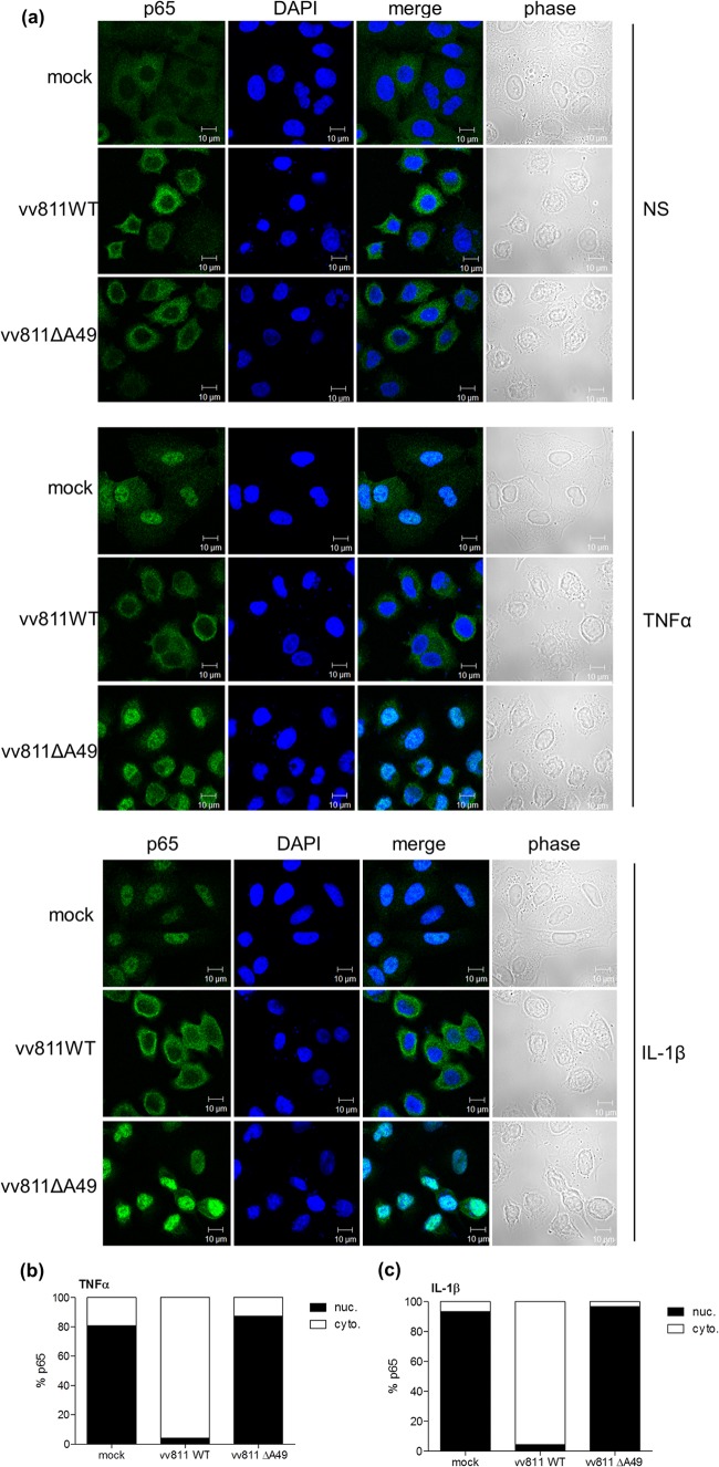
FIG 3.
vv811ΔA49 does not inhibit p65 translocation in response to TNF-α or IL-1β stimulation. (a) A549 cells were mock infected or infected for 6 h with 5 PFU per cell of the indicated viruses and then stimulated for 30 min with TNF-α (50 ng/ml) or IL-1β (12.5 ng/ml) or nonstimulated (NS) as a control by incubation with medium alone. The cells were fixed and stained for immunofluorescence analysis using an antibody against intracellular p65 (green). Nuclei and viral factories were visualized by DAPI staining (blue). Merged and phase-contrast images are also shown. (b, c) The percentage of cells in which p65 was either cytoplasmic (cyto.) or nuclear (nuc.) was calculated from 250 to 300 cells for each condition in panel a.

