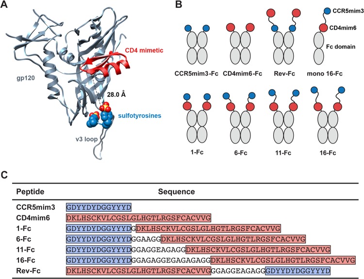
FIG 1.
Receptor-mimetic constructs. (A) A ribbon diagram of HIV-1 gp120 (gray) is shown bound to a CD4-mimetic peptide (red) and to a sulfopeptide (blue, with sulfate oxygen and sulfur atoms shown in red and yellow, respectively). Sulfotyrosines were derived from the heavy chain of CD4-induced antibody 412d. The figure was generated by aligning the gp120 molecules of the gp120-CD4-412d complex (Protein Data Bank [PDB] 2QAD) with those of the gp120-F23 complex (PDB 1YYM) (10, 19). The dashed line indicates the 28-Å distance from the amino-terminal α-carbon of the CD4 mimetic to the α-carbon of 412d sulfotyrosine 100. (B) Diagram of mimetic-Fc fusion constructs studied here. CCR5mim3 is shown as a blue circle and CD4mim6 as a red circle, and the Fc domain (including the hinge, CH2, and CH3 domains of human IgG1) is indicated in gray. (C) Sequences of the single- and double-mimetic peptides used in this paper. Blue indicates CCR5mim3, and red indicates CD4mim6. Linker regions are not highlighted.

