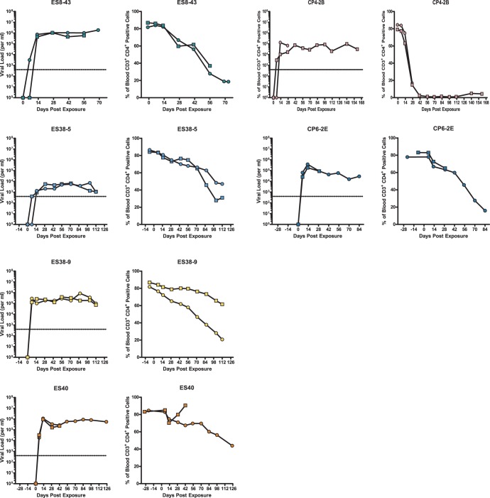
FIG 4.
In vivo replication and pathogenesis of HIV isolates from ES and CPs. BLT humanized mice were exposed to HIV-1 isolates that were cultured from either ES or CPs. Each isolate was used to infect two mice. The mice were bled periodically to obtain plasma for viral load analysis via real-time RT-PCR and blood mononuclear cells for flow cytometric analysis. For each isolate, the left panel shows viral load analysis (the dotted line represents the limit of detection for the viral load assay), and the right panel shows the percentage of peripheral blood CD4+ T cells. Different symbols represent different mice. One mouse infected with isolate CP4-2B and one mouse infected with isolate ES40 died shortly after day 28 and day 42 of infection, respectively.

