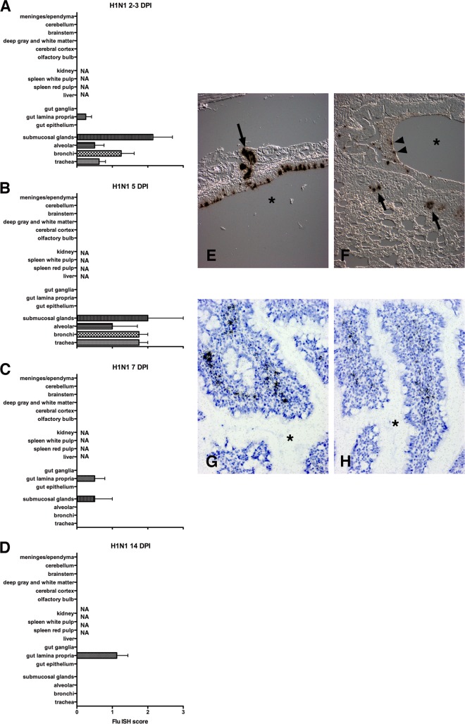
FIG 1.
Histograms delineate the average ISH score for different regions of organs 2 to 3 (A), 5 (B), 7 (C), and 14 (D) days after primary H1N1pdm09 infection. In the first 5 days of infection, the majority of the viral infection was limited to lung with predominant infection of bronchial epithelium and submucosal glands. By 7 DPI most of the lung infection had cleared except for occasional submucosal glands. A mild focal infection in the small bowel was limited to the lamina propria. NA, tissue not available. (E) ISH on paraffin section from trachea at 5 DPI. ISH demonstrates influenza virus RNA (dark grains) in tracheal epithelium and submucosal glands (arrow). The asterisk is in the tracheal lumen. (Differential interference contrast [DIC] without counterstain.) (F) ISH on paraffin section from lung at 5 DPI demonstrates influenza virus RNA in bronchial epithelium, necrotic debris (arrowheads), and submucosal glands (arrows). The asterisk is in the bronchial lumen. (DIC without counterstain.) (G and H) ISH on paraffin sections from small bowel at 14 DPI demonstrates influenza virus RNA (dark grains) in lamina propria. The asterisk is in the bowel lumen between villi. For panels G and H, tissues were counterstained with hematoxylin. Scoring: 0 = no definitive signal, 1 = occasional focus, 2 = focus in most fields, 3 = more than one focus per field.

