FIG 4.
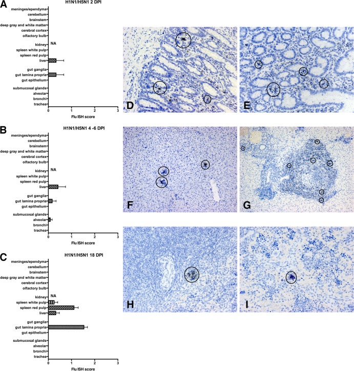
Ferrets were initially infected with H1N1pdm09 (Mex/09) and then 2 to 3 months later challenged with H5N1 as described in the text. Histograms delineate the average ISH score for different regions of organs at 2 (A), 4 to 6 (B), and 18 (C) days after H5N1 challenge. In the first 2 days after challenge, influenza virus RNA is limited to small bowel lamina propria and regions of periportal hepatitis. At 4 to 6 DPI, ISH demonstrates little viral infection, which is limited to liver, lamina propria, and occasional alveolar cells. At 18 DPI, ISH demonstrates limited viral RNA in hematopoietic elements of the spleen, liver, and lamina propria. At 6 (D) and 18 (E) DPI, infected cells (circled) can be detected focally in the small bowel lamina propria. At 2 (F) and 18 (G) DPI, infected cells (circled) can be seen in liver in regions of periportal hepatitis. At 18 DPI, infected cells (circled) can be detected in splenic white (H) and red (I) pulp. All ISH slides were counterstained with hematoxylin. Scoring: 0 = no definitive signal, 1 = occasional focus, 2 = focus in most fields, 3 = more than one focus per field.

