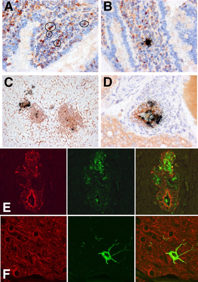
FIG 6.
To better define the lineages of infected cells, we stained paraffin sections with cell-specific markers in conjunction with ISH for influenza viral RNA. (A to C) Sections stained with Griffonia simplicifolia lectin that binds to ferret macrophages. (D) Section stained with anti-cytokeratin antibody that binds to epithelial cells. (A and B) Small bowel sections show infected cells (circled) in the lamina propria that display both peroxidase reaction product (red) and ISH grains (black), indicating infection of lamina propria macrophages. (C) Liver section demonstrates infected macrophage elements in regions of periportal hepatitis. (D) Some bile duct epithelial cells labeled for cytokeratin (red) also hybridize with the influenza virus probe (black grains). (E and F) Double-label immunofluorescent images illustrate inflamed liver bile duct epithelia stained for cytokeratin (red) that colocalize with H5 HA protein (green) (E) and infected neurons that stain with microtubule-associated protein-2 (red) and influenza virus H5 HA protein (green) (F).

