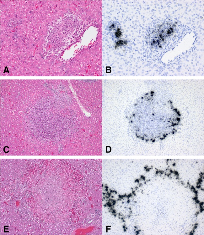
FIG 7.
Ferrets supplied for these experiments had a low-grade chronic hepatitis independent of influenza virus infection. With H5N1 challenge, animals showed 3 patterns of influenza-related hepatitis: acute periportal duct inflammation (A and B), severe chronic inflammatory nodules (C and D), and multifocal hepatic necrosis (E and F). In the first pattern, a hematoxylin and eosin (H&E)-stained section of the liver (A) shows acute bile duct inflammation, while ISH on a sequential section (B) demonstrates severe H5N1 infection of inflamed periportal ducts. In the second pattern, an H&E-stained section (C) illustrates a large nodule of chronic inflammatory cells that on ISH (D) shows numerous H5N1-infected cells both in the center and at the perimeter. An H&E-stained section (E) of the third pattern shows central necrosis with ISH (F) demonstrating a perimeter of infected cells surrounding the central necrosis.

