
FIG 1.
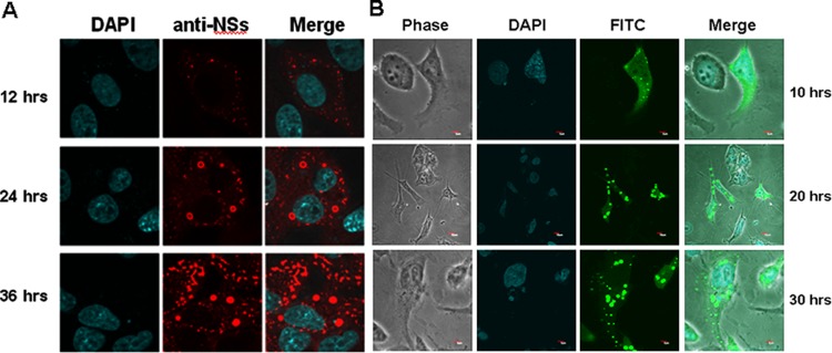
Inclusion bodies (IBs) formed by NSs in infected and transfected cells. (A) Vero cells were infected with SFTSV at an MOI of 1, and the cells were fixed and permeabilized at the indicated time points. The cells were stained with a rabbit anti-NSs antibody at 1:20 dilution in PBS, followed by staining with a TRITC-conjugated anti-rabbit IgG, and subjected to confocal microscopy (magnification, ×400). (B) A cDNA of NSs was cloned into pEGFP, and the plasmid was used to transfect HeLa cells. Transfected cells were subjected to confocal microscopy at the indicated time points.