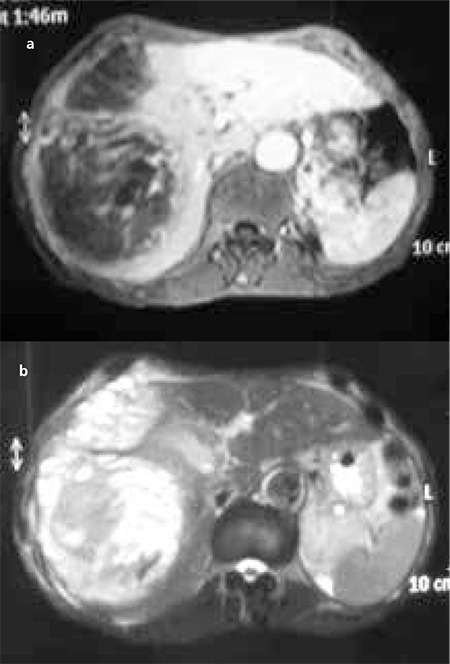
Figure 1. Magnetic resonance imaging. There was a large mass (152x120x135 mm) with heterogeneously hypointense on the T1-weighted image (a), and heterogeneously hyperintense on the T2-weighted image. After the administration of contrast material, heterogeneous hyperenhancement was observed in the solid parts of the lesion (b). This mass covered segment 7-8 completely and segment 4 partially.

