
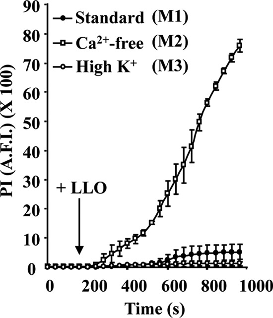
FIG 2.
Host cells damaged by LLO undergo Ca2+-dependent but K+-independent membrane resealing. Perforation of HepG2 cells was measured by quantitative live-cell fluorescence microscopy. Cells were incubated on the microscope stage at 37°C for 980 s with 20 μM PI in standard medium (M1), Ca2+-free medium (M2), or high-K+ medium (M3). Phase-contrast and fluorescence images were recorded at regular time intervals using a 40× objective, and 1 nM LLO was added after 160 s of incubation. Results are expressed as the average fluorescence intensity (A.F.I.) of the cell area ± SEM. By 600 s, there was a statistically significant difference between the levels of PI incorporation in cells incubated in M1 and in M2. There was no significant difference between samples incubated in M1 and M3 at any time point.