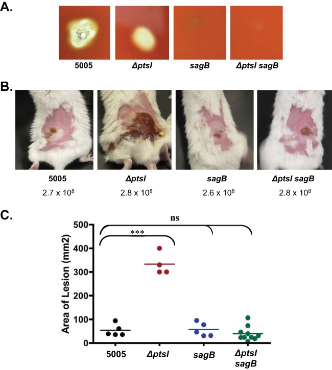
FIG 5.
Role of streptolysin S in ΔptsI increased lesion formation. (A) Zones of hemolysis for wild-type (WT) MGAS5005, ΔptsI single mutant, ΔsagB single mutant, and ΔsagB ΔptsI double mutant strains on 5% sheep blood agar plates after growth at 37°C. (B) Representative images of mice infected s.c. with WT MGAS5005, MGAS5005.ΔptsI mutant, ΔsagB single mutant, and ΔptsI sagB double mutant strains. The numbers of CFU used in infection are indicated. (C) Lesion size of the same experiment measured at 38 h after s.c. infection of MGAS5005 (black), ΔptsI mutant (red), ΔsagB mutant (blue), and ΔptsI sagB double mutant (green). Data represent two independent experiments, and significance was determined using the unpaired two-tailed t test (***, P ≤ 0.001; ns, not significant).

