
FIG 1.
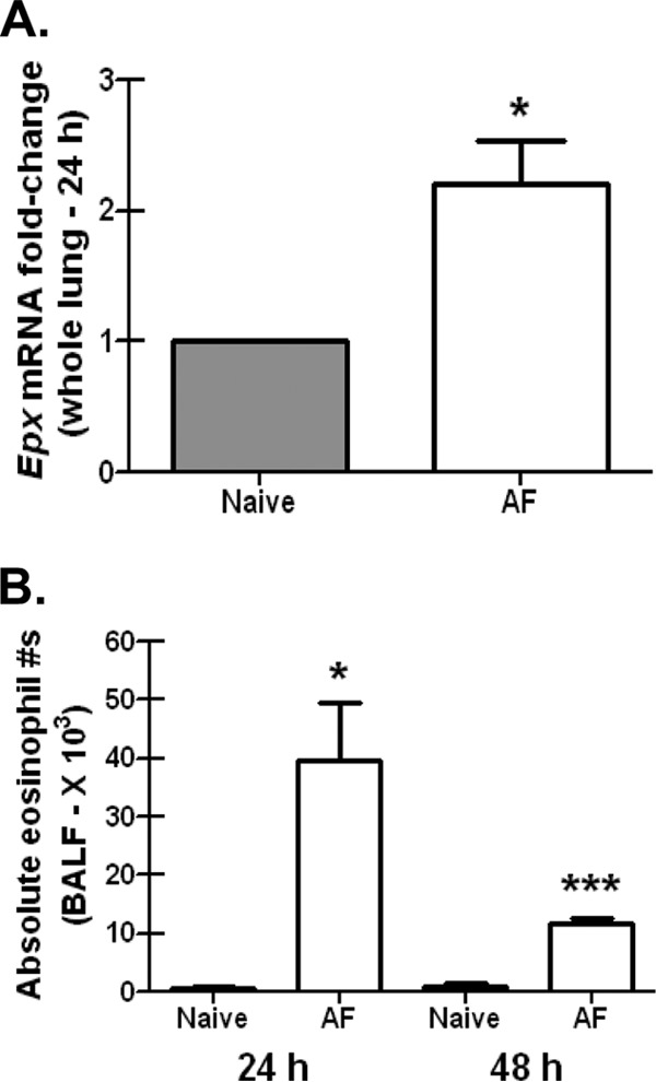
Eosinophils are recruited to lung after A. fumigatus challenge. (A) BALB/c wild-type mice were challenged intratracheally with 7 × 10⁷ A. fumigatus conidia (AF), and 24 h postexposure, whole lungs were collected, total RNA was isolated and transcribed to cDNA, and quantitative real-time PCR was performed for Epx. Gene expression was normalized to Gapdh, and fold changes between naive mice (set at 1) and A. fumigatus-exposed BALB/c mice were determined using the 2−ΔΔCT method. Cumulative data from three independent studies (n = 3 to 5 mice per group per study) are illustrated. *, P value of <0.05 (paired two-tailed Student t test). (B) BALB/c wild-type mice were challenged intratracheally with 7 × 10⁷ A. fumigatus conidia, and 24 or 48 h postexposure, lung cells were isolated via bronchoalveolar lavage, Fc blocked, stained with a LIVE/DEAD staining kit, and thereafter stained with fluorochrome-conjugated CD11c, CD11b, Gr-1, and Siglec F. Cumulative data from three independent studies (n = 2 or 3 mice per group per time point per study) are illustrated. Data are expressed as absolute numbers of live cells in lung lavage fluid. * and ***, P values of <0.05 and <0.0001, respectively (unpaired two-tailed Student t test).