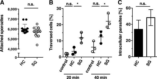
FIG 1.

Hemocoel sporozoites display a distinct impairment of cell traversal. (A) Sporozoite adhesion to hepatoma cells. Hepatoma cells were incubated with hemocoel sporozoites (black circles; HC) or salivary gland sporozoites (white circles; SG), and adherent sporozoites were quantified. Each dot represents one sample. Shown are mean values (± the standard deviation [SD]) from three independent experiments. (B) Sporozoite traversal of hepatoma cells. Hepatoma cells were incubated with FITC-dextran either alone (white circles; control), with hemocoel sporozoites (black circles; HC), or with salivary gland sporozoites (gray circles; SG) for 20 and 40 min. Cells were fixed and analyzed by FACS to enumerate the percentage of dextran-positive cells. The results represent mean values (± the SD) of three independent experiments with three samples each. (C) Sporozoite invasion of hepatoma cells. Hepatoma cells were infected with hemocoel sporozoites (black bar, HC) or salivary gland sporozoites (white bar, SG) for 2 h. Cells were fixed and extracellular sporozoites were stained with an anti-CSP antibody, followed by permeabilization and staining with an anti-GFP antibody, in order to distinguish intracellular parasites that have invaded the cell versus attached parasites. The results represent mean values (± the SD) of three independent experiments with two samples each. n.s., not significant; *, P < 0.05 (unpaired t test).
