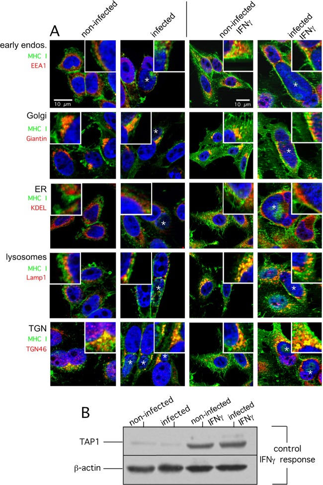
FIG 3.
MHC I subcellular localization in C. trachomatis serovar D-infected HeLa cells. (A) HeLa cells were infected for 48 h or not infected (columns 1 and 2) and costained for MHC I (green) and EEA1 (early endosomes), Giantin (Golgi compartment), KDEL proteins (ER), LAMP1 (lysosomes), and TGN46 (trans-Golgi compartment network) (red). Cells were infected with Chlamydia or not infected as in columns 1 and 2 and additionally treated with IFN-γ (columns 3 and 4). In particular, cells were treated with IFN-γ for 48 h prior to infection with Chlamydia and for 48 h during infection with Chlamydia. (B) The proper IFN-γ response of infected and noninfected cells was controlled via the cytokine-mediated induction of TAP1 analyzed by corresponding Western blot assays. The corresponding overlays of the immunostainings are depicted. The insets show magnifications of representative cell areas where MHC I and organelle markers colocalized. For direct comparison, all cells were photographed with the same exposure time. DNA of the host nucleus and chlamydial inclusions were labeled with 4′,6-diamidino-2-phenylindole (blue). Parasitophorous vacuoles are indicated by asterisks.

