FIG 4.
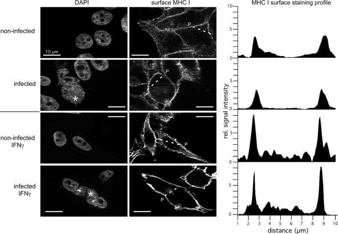
MHC I localization on the plasma membrane of infected and noninfected HeLa cells in the absence or presence of IFN-γ. HeLa cells were pretreated with IFN-γ for 48 h and also during the following 48 h of chlamydial infection. The proper IFN-γ response of infected and noninfected cells was controlled via cytokine-mediated TAP1 induction in Western blot assays (not shown). Nonpermeabilized cells were immunostained for surface MHC I (right panel). DNA of the host nucleus and inclusions of the same cells were labeled with 4′,6-diamidino-2-phenylindole (left panel). Parasitophorous vacuoles are indicated by asterisks. Noninfected and infected cells were photographed with the same exposure time. To compare MHC I surface expression in infected and noninfected HeLa cells, we measured the intensity of transmitted fluorescent light along a line segment drawn across the cellular area of interest (indicated by a broken line and P). The representative fluorescence intensity profiles measured across the cells along the dashed lines are shown on the right. rel., relative.

