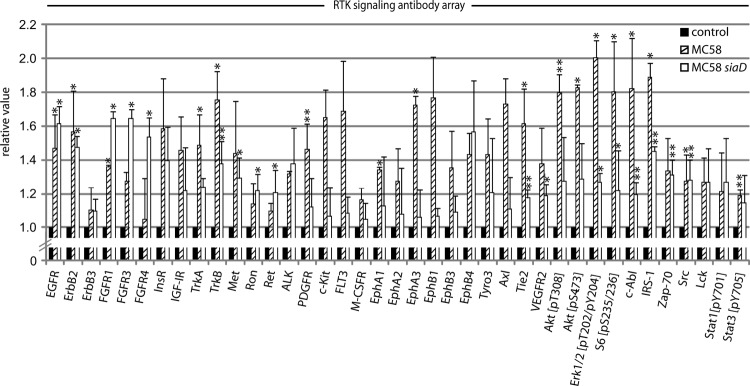
FIG 1.

Evidence for phosphorylation of RTKs and key signal proteins in N. meningitidis-infected HBMEC. The PathScan RTK signaling antibody array kit was utilized to analyze the phosphorylated signal levels of kinases and key signaling nodes (see Materials and Methods). HBMEC were infected with MC58 or MC58 siaD at an MOI of 30 or were left uninfected (control), and cell lysates were collected after 3 h. Dot densities were analyzed with ImageJ software. The values for control cells were set as 1, and the values of MC58 and MC58 siaD are shown relative to the control. Results represent mean values ± standard deviations of three independent experiments. *, P < 0.05; **, P < 0.01 relative to untreated cells.
