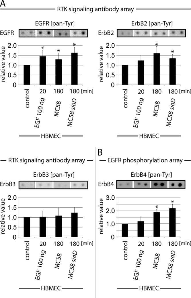
FIG 2.

N. meningitidis infection causes activation of EGFR, ErbB2, and ErbB4. HBMEC either were left uninfected (control) or were incubated for 20 min with 100 ng/ml EGF (positive control) or infected for 180 min with N. meningitidis strain MC58 or MC58 siaD, and then cell lysates were analyzed using the PathScan RTK signaling antibody array (A) or the human EGFR phosphorylation antibody array (B) as described in Materials and Methods. A chemiluminescent film image (upper panel) and the quantification of that image (lower panel) are shown. The chemiluminescent array images were captured following 60-s film exposures. Dots were quantified by densitometric analysis using ImageJ. Graphs represent data from three independent experiments. *, P < 0.05.
