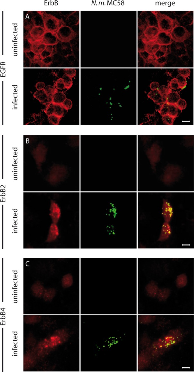
FIG 3.

EGFR, ErbB2, and ErbB4 accumulated at the site of meningococcal adhesion. HBMEC were infected with N. meningitidis at an MOI of 30 for 1 h, fixed, and examined by confocal microscopy. Bacteria were stained with a rabbit antimeningococcal serum and counterstained with an anti-rabbit Cy3 antibody (green). ErbB receptors were stained with either anti-EGFR (A), anti-ErbB2 (B), or anti-ErbB4 (C) followed by counterstaining with anti-mouse Cy5 antibody (red). Uninfected HBMEC served as controls. Optical sections were acquired using a confocal microscope (Leica SP5). The figure shows representative images of single optical sections from three independent experiments. Bar, 5 μm.
