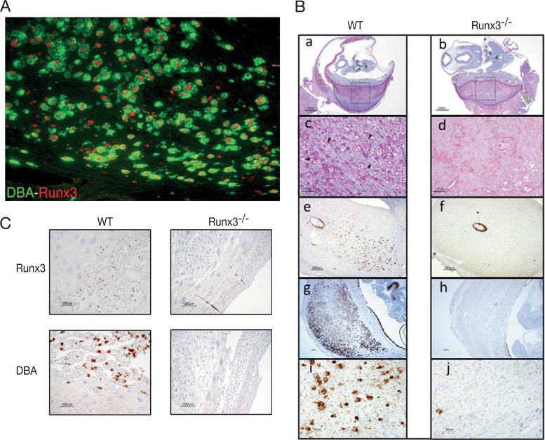
FIG 4.
Runx3−/− pregnant mice lack uNKC. (A) DBA and anti-Runx3 immunostaining show Runx3 expression in uNKC. (B) WT and Runx3−/− implantation site sections stained with PAS (a to d, E11; panels c and d represent enlarged boxed regions in panels a and b, respectively; arrowheads in panel c mark uNKC) or DBA (e and f, E6.5; g to j, E11; panels i and j represent enlarged regions in panels g and h, respectively). (C) IHC of Runx3 (upper panels) and DBA (lower panels) on sections of E10.5 implantation sites of pregnant Rag−/− γc−/− mice to which WT (left panels) or Runx3−/− (right panels) fetal liver cells were transferred ∼2 months earlier. Bars, 1 mm (Ba and b), 50 μm (Bc, d, i, and j), 250 μm (Be to h), and 100 μm (C).

