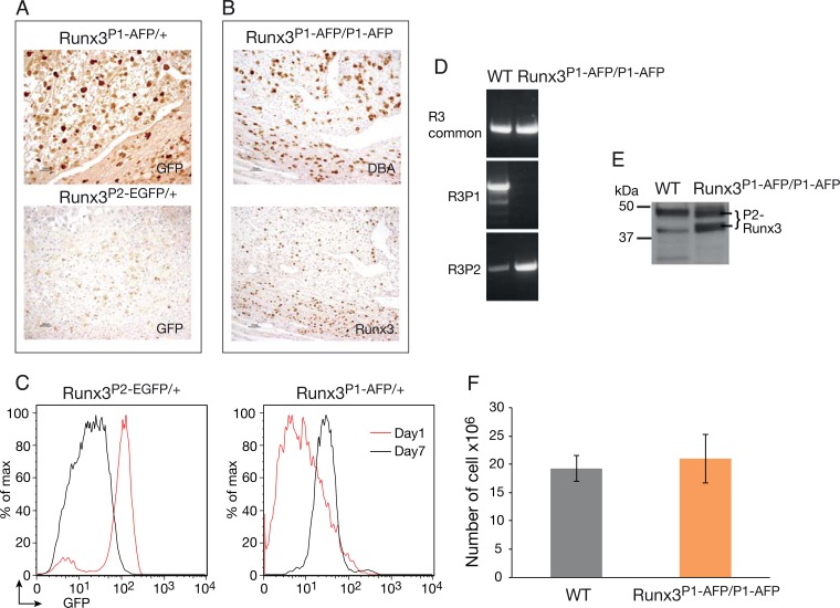
FIG 5.
Runx3 promoter usage in NKC. (A) E11 implantation site sections of Runx3P1-AFP/+ or Runx3P2-EGFP/+ pregnant mice stained with anti-GFP. (B) E11 implantation sites of Runx3P1-AFP/P1-AFP mice analyzed by DBA IHC (upper panel) or Runx3 (lower panel). Bars (A and B), 50 μm. (C) FACS analysis of GFP expression in IL-2-cultured NKC of Runx3P1-AFP/+ and Runx3P2-EGFP/+ mice. (D) RT-PCR analysis of Runx3 common and P1- and P2-specific transcripts in RNA prepared from WT and Runx3P1-AFP/P1-AFP NKC on day 7 of culture with IL-2. (E) Western blot documenting Runx3 expression in IL-2-cultured WT and Runx3P1-AFP/P1-AFP NKC. The 46-kDa Runx3 P1 isoform is clearly detected in the WT lane, while in the Runx3P1-AFP/P1-AFP lane, the two typical P2 Runx3 45- and 43-kDa isoforms (76) are detected. (F) Analysis of WT and Runx3P1-AFP/P1-AFP NKC numbers following 7 days in culture with IL-2.

