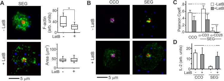
FIG 3.
Increased mobility of membrane proteins allows primary human T cells to respond to segregated patterns. (A) LatB wash-in disrupts the IS cytoskeleton and decreases localization of pSFK to OKT3-containing features on micropatterned surfaces. These images illustrate primary human T cells 30 min after seeding on a SEG patterned surface and show anti-CD3 (red), anti-CD28 (blue), and F-actin (green). The graphs quantitatively compare per-area actin intensity and cell spreading. The data are from a representative experiment (n > 20 cells per surface). The data were compared using Kruskal-Wallis methods (*, P < 0.005). (B) LatB wash-in also changed the distribution of pSFK (green) within these cells. These images illustrate cells 30 min after seeding on CCO and SEG patterns. Areas of anti-CD3 and anti-CD28 are shown in red and blue, respectively, while colocalized patterns appear in purple. (C) Wash-in of LatB changes pLck correlation with anti-CD3 and anti-CD28. The data represent means ± the SD for 10 to 15 cells on each pattern. The data were analyzed using ANOVA/Tukey methods (α = 0.05). (D) IL-2 secretion on segregated but not colocalized patterns is enhanced by application of LatB. The data are means ± the SD from three experiments and were analyzed using ANOVA/Tukey methods (α = 0.05).

