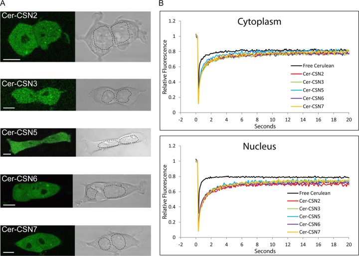
FIG 1.
Cellular CSN is localized in the cytoplasm and nucleus and displays the mobility of a large macromolecular complex. (A) Representative fluorescent midsection images of live HEK293 cells, stably transfected with CSN subunits N-terminally fused to Cerulean. The images indicate that CSN subunits are localized in the cytosol and nucleus but are excluded from nucleoli. For convenience, the nuclear borders are marked with a dashed line. Scale bars represent 10 μm. (B) FRAP curves of fluorescent CSN subunits and free Cerulean. Each plot constitutes an average of at least 60 cells, normalized to prebleach intensity.

