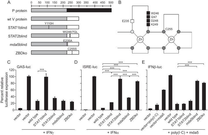
FIG 2.
Generation of CDV V-protein mutants unable to interfere with different IFN signaling pathways. (A) Overview of mutations introduced in the P gene to produce V proteins that are unable to interfere with STAT1, STAT2, or mda5 signaling or that have a disrupted zinc-binding domain (ZBDko). The P/V shared domain is shown in light gray, the RNA editing site is indicated by a black line, and the unique regions of the P or V protein are shown in dark gray or white, respectively. wt, wild type.(B) Positions of the respective mutations in the zinc finger structure of the unique V-protein carboxy terminus (adapted from reference 11 with permission of the publisher). Conserved cysteines (dark gray filled circles) and the conserved histidine (light gray filled circle) are indicated. The mda5-interacting residue E235 (white box) and the STAT2-interacting residues (black boxes) are shown. (C to E) IFN signaling interference activity of the V-protein mutants. Huh7 cells were transfected with either a GAS-luciferase reporter plasmid (C) or ISRE-luciferase reporter plasmid (D) or an IFN-β-luciferase reporter gene plasmid (E) together with a mda5 expression plasmid, in combination with either an empty vector, or vectors expressing the CDV wild-type V protein, or the respective mutant CDV V proteins as indicated. Cells were treated with 100 ng/ml of IFN-γ or 1,000 U/ml of IFN-α or transfected with 1 μg/ml of poly(I·C) for 18 h, 8 h, or 20 h prior to lysis, respectively, and luciferase activity was measured. Luciferase activity of cells transfected with an empty vector was set at 100%. Values are the averages of three experiments done in triplicate; error bars indicate the standard errors of the means (SEM). Asterisks show the levels of statistical significance for the values indicated by the ends of the bars (***, P < 0.001).

